Prostate cancer
Prostate cancer occurs in tissues of the prostate. The main signs of prostate cancer include a weak urine flow or frequent urination. Urine and blood tests are used to detect prostate cancer. The prostate is a gland in the male reproductive system. It lies just below the bladder(the organ that collects and empties urine) and in front of the rectum (the lower part of the intestine). It is about the size of a walnut and surrounds part of the urethra (the tube that empties urine from the bladder). The prostate gland makes fluid that is part of the semen.
The prostate is the gland below a man’s bladder that produces fluid for semen. Prostate cancer is common among older men. It is rare in men younger than 40. Risk factors for developing prostate cancer include being over 65 years of age, having a family history, and being African-American.
Symptoms of prostate cancer may include Problems passing urine, such as pain, difficulty starting or stopping the stream or dribbling, Low back pain, and pain with ejaculation.
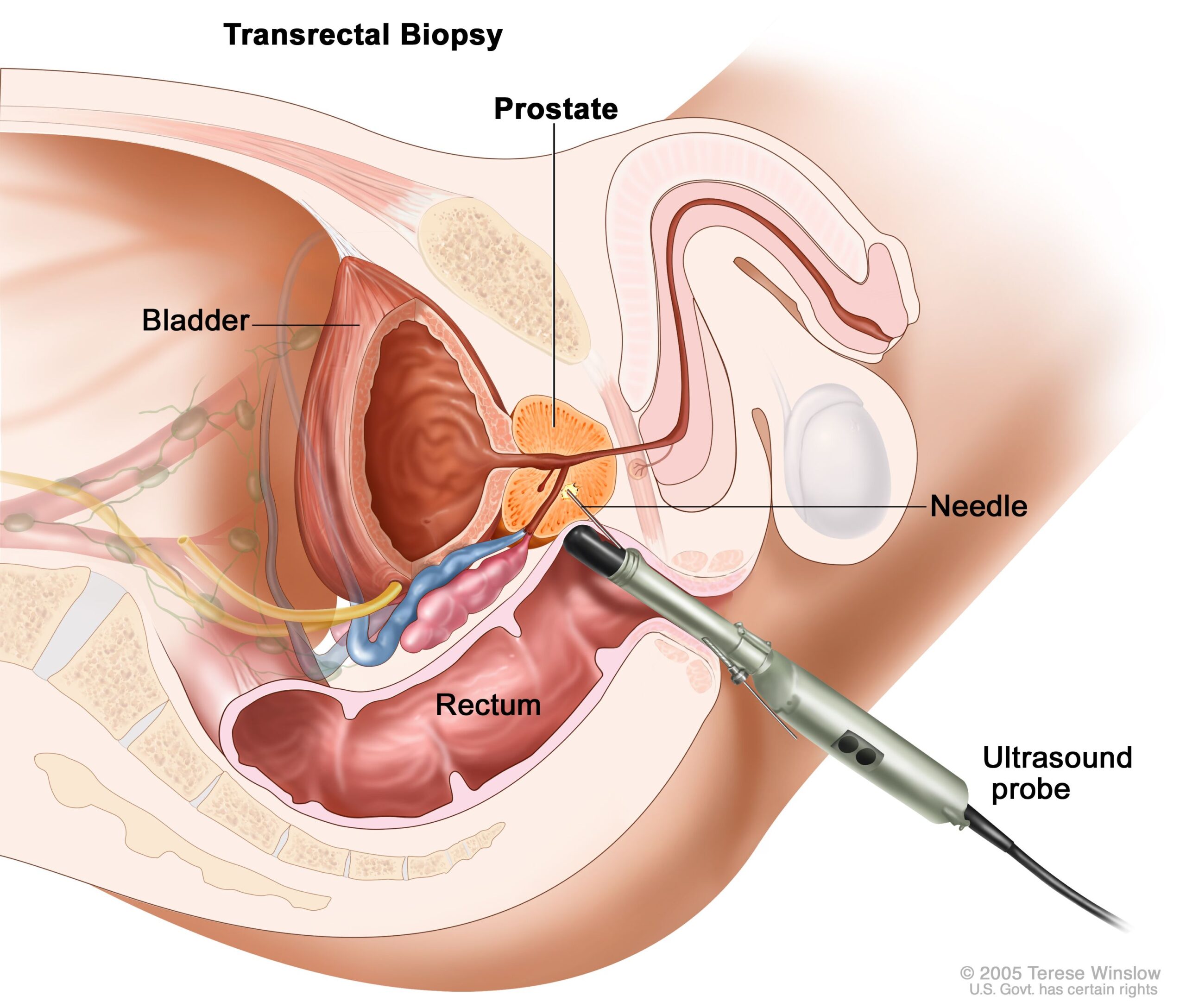
To diagnose prostate cancer, a digital rectal exam to feel the prostate for lumps or anything unusual may be done by a practitioner. You may also get a blood test for prostate-specific antigen (PSA). These tests are also used in prostate cancer screening, which looks for cancer before you have symptoms. If your results are abnormal, you may need more tests, such as an ultrasound, MRI, or biopsy.
Treatment often depends on the stage of cancer. How fast cancer grows and how different it is from surrounding tissue helps determine the stage. Men with prostate cancer have many treatment options. The treatment for one man may not be best for another. The options include watchful waiting, surgery, radiation therapy, hormone therapy, and chemotherapy. You may have a combination of treatments.
Prostate cancer is the second most common cause of male cancer deaths and will afflict 1 in 6 men. Many diagnostic prostate biopsies fail to detect occult cancers because of the difficulty in adequately determining the prostate’s anatomy. Approximately 20% of ultrasound image-guided prostate biopsies return negative results despite rising levels of prostate-specific antigen (PSA) markers in the blood and the presence of cancer somewhere in the 30+ cc gland. These failures have been attributed by some to the hesitation to sample one of the two major parts of the glands— namely, the predilection toward the more easily reached peripheral zone (PZ) over the less accessible central gland (CG). We can outline these two non-overlapping adjacent regions of the gland (see image).
Advancing solutions to this vexing clinical problem will raise awareness about the impact computer image processing can have on addressing a widespread, critical healthcare puzzle. Open and closed source algorithms applicable to both 1.5T and 3T magnetic resonance images (MRIs) will be most welcome.